|
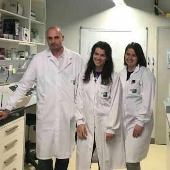
USC researchers (Photo: courtesy of USC)
An investigation led by the USC discovers a new case of cancer that is spread between species such as the clam
 SPAIN
SPAIN
Friday, January 21, 2022, 07:00 (GMT + 9)
A project involving researchers from the University of Santiago de Compostela (USC) opens a new path for the study of cancer in humans.
The work, published in the journal eLife, shows how cancer cells pass from one bivalve to another.
"It is a new point of view to understand metastasis, the spread of cancer to other organs of the body," they say.
Clams can spread a type of cancer similar to leukemia in humans. The disease is transmitted between bivalves as a virus. It goes from sick specimens to healthy ones. Therefore, it survives the death of the clam that originated it. This has been confirmed by a study in which researchers from the Singular Center for Research in Molecular Medicine and Chronic Diseases (CiMUS) of the University of Santiago de Compostela have participated.
.jpg)
Universidad de Santiago de Compostela (USC)
The research, published in the journal eLife, not only reveals new data on the little-known contagious nature of cancer. Also, these scientists say, it opens a new avenue for the study of cancer in humans.
“The genetic study of these contagious cancers is a new point of view to try to understand metastasis, that is, the spread of cancer to other organs of the body. With this new case of contagious cancer, we now have more models to study the genetic causes of cancer transmissibility”, explains Professor José Tubío, the scientist leading the research.
500 units of clam
.jpg) <------ Foto: Universidad de Santiago de Compostela (USC) <------ Foto: Universidad de Santiago de Compostela (USC)
To carry out the study, the researchers collected more than 500 clam units. Bivalves that came not only from the coast of Galicia, but also from Ireland, France, Croatia and Portugal, countries that also participate in the European project called Scuba Cancers.
During the investigations, the scientists managed to find a type of cancer similar to leukemia in humans, called hemic neoplasia. DNA sequencing technologies made it possible to discover that cancer cells of the original species could "jump between species behaving in an infectious manner", points out Alicia L. Bruzos, one of the CiMUS researchers who has participated.
Contagious cancers were only recently discovered thanks to advances in the field of genetics. These have made it possible to determine in which individual a tumor cell originated.
.jpg)
Geographical location of warty venus (V. verrucosa) specimens and diagnosis of hemic neoplasia.
(a) Locations of V. verrucosa clams collected for this study and specimens diagnosed with hemic neoplasia. Size of the pie charts correlates with the number of samples collected (number of samples ‘n’ is shown together with each pie chart). Pie charts show the proportion of samples with hemic neoplasia (black, no neoplastic specimens; red, neoplastic specimens). Codes of neoplastic samples are shown. Top-right corner shows a representative specimen of the species V. verrucosa. (b) Cytological examination of haemolymph smear (Hemacolor stain) from a healthy (N0) specimen, ERVV17-2963, shows normal haemocytes. (c) Haemolymph smear of a V. verrucosa specimen with high-grade (N3 stage) hemic neoplasia, ERVV17-3193, shows neoplastic cells that replaced normal haemocytes. (d) Detail of haematoxylin and eosin-stained of histological section from the gills of the healthy (N0) specimen ERVV17-2990. (e) Same for ERVV17-2995, a specimen infected with a high-grade (N3 stage) hemic neoplasia, showing neoplastic cells infiltrating the gills. (f) Transmission electron microscopy analysis of a V. verrucosa hemic neoplasia tumour cell shows a round shape, pseudopodia ‘p’, pleomorphic nucleus ‘n’ with scattered heterochromatin, and mitochondria ‘m’. (g) Metaphase chromosomes from a neoplastic cell found in the gills of the V. verrucosa specimen EVVV11-02, showing abnormal chromosome number (>19 pairs) and abnormal chromosome morphology. Chromosomes stained with 4′,6-DiAmidino-2-PhenylIndole (DAPI) and Propidium Iodide (PI). Image: Paper/Universidad de Santiago de Compostela (USC)
Contagious cancers are currently only known in dogs, Tasmanian devils, and various species of mussels, clams, and cockles. The study of DNA, both nuclear and mitochondrial, revealed, as evidenced in the article, that this cancer originated in a different donor clam in which it inhabits the same regions as the first recipient clams.
An ecological threat
The spread of cancer between closely related species warns of the danger posed by these contagious cancers. "Our discoveries confirm that transmissible marine cancers can jump between species and point to the need for their identification and genetic characterization for their prevention because it could pose a danger to the marine ecosystem and a serious ecological threat," says this CiMUS researcher.
.jpg)
Mitochondrial DNA sequencing and phylogenetic analyses reveal cancer contagion between warty venus (V. verrucosa) and striped venus (C. gallina) clam species.
(a) In eight warty venus specimens sequenced with Illumina paired-ends, the pie charts show the proportion of reads mapping Cox1 reference sequences from 137 different Verenidae species, including V. verrucosa (red), C. gallina (blue), and the remaining species (grey). Two different tissues were sequenced: the tumour tissue (left pie chart), typically haemolymph, and the host/matched-normal tissue (right pie chart), typically foot. Note that for specimen EVVV11-02 only the host/matched-normal tissue (gills) was available. ‘n’ denotes the total number of reads mapping the Cox1 reference for the tumour tissue (left), and the host tissue (right). (b) Representative specimen of the species C. gallina. (c) Capillary sequencing electropherograms of mitochondrial Cox1 gene fragments from two neoplastic V. verrucosa specimens (EMVV18-373 and EVVV11-02) and two healthy reference specimens from V. verrucosa and C. gallina. The results show overlapping peaks (arrows) in the sequenced tissues from the neoplastic animals, which suggest coexistence of mitochondrial DNA (mtDNA) haplotypes from two clam species. (d) In V. verrucosa neoplastic (N2-stage) specimen EMVV18-400, mtDNA read depth shows different proportion of warty venus and striped venus mtDNA haplotypes in the tumour tissue (haemolymph) and the matched-normal tissue (foot). (e) Molecular phylogeny using Bayesian inference inferred on the alignment of all mitochondria coding genes and rRNA gene sequences (15 loci) that includes six neoplastic V. verrucosa specimens with evidence of cancer contagion from C. gallina. Bootstrap values are shown above the branches.Image: Paper/Universidad de Santiago de Compostela (USC)
"A previous study on the contagious cancer of mussels found on the European and South American coasts proposed travel between mussels attached to the cases of ships that made commercial trips between Europe and South America as a potential route of contagion," says Alicia L. Bruzos . This suggests deepening the knowledge of the complete route of cancer contagion, and the routes of transport to know the real causes of transmission.
Contagious canker survives the death of the clam in which it originated by infecting new clams. In fact, the contagious cancer that dogs get was estimated to be about 8,500 years old. "Future studies will allow us to shed light on the age of the contagious cancer of the donor clams, but, at a minimum, they are known to be about nine years old, since the samples analyzed in this study were taken between 2011 and 2020," he says. the researcher Daniel García-Souto.
.jpg)
Nuclear DNA sequencing and phylogenetic analyses confirm a single cancer lineage spreading in populations of the warty venus (V. verrucosa) that originated in the striped venus (C. gallina).
(a) Single-nucleotide variants discriminating between V. verrucosa tumours and the three canonical species (V. verrucosa, C. gallina, and C. striatula) along a 441- and a 559-bp long fragments of nuclear genes DEAH12 and TFIIH, respectively. (b) Maximum Likelihood molecular phylogenies based on the two fragments of the nuclear DNA markers DEAH12 and TFIIH. Bootstrap support values (500 replicates) from Maximum Likelihood analyses above 50 are shown on the corresponding branches. (c) Multispecies coalescent (MSC) tree of V. verrucosa, their tumours and Chamelea sp. based on the entire mitochondrial DNA (mtDNA) and the two nuclear markers, DEAH12 and TFIIH. A maximum clade credibility (MCC) tree is shown, with posterior probabilities below the branches, and 95% highest probability density (HPD) intervals of node heights as grey bars. The trees distribution shown includes 1000 trees and represents the range of alternative topologies, in which blue is the most common set of topologies, red the second most common one, and green the remaining. (d) Fluorescence in situ hybridization (FISH) to specifically detect the satellite DNA CL4 in one V. verrucosa tumour and healthy specimens from the species C. gallina and V. verrucosa shows probes accumulate in heterochromatic regions, mainly in subcentromeric and subtelomeric positions, from the chromosomes of the tumour and the healthy C. gallina tested but not in healthy V. verrucosa. Image: Paper/Universidad de Santiago de Compostela (USC)
The Scuba Cancers project is funded by the European Research Council (ERC) with 1.5 million euros.
Editado por Malena Nahum
[email protected]
www.seafood.media
|


 Print
Print